|
|
|
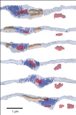
Three-dimensional reconstruction of granule
cell presynaptic boutons. In this
example six reconstructed granule cell
presynaptic boutons are shown, with mitochondria
indicated in tan, vesicles indicated by blue and
the postsynaptic density indicated in red.
Associated with each bouton is the postsynaptic
density and the position of the morphologically
docked vesicles that are well positioned next to
the plasma membrane. |
|
|
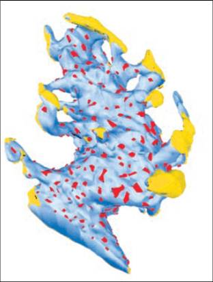
Three-dimensional reconstruction of a mossy
fiber presynaptic bouton. Red regions
indicate postsynaptic densities where the mossy
fiber forms synaptic contacts onto granule cell
dendrites. The yellow regions are in
contact with glia.
|
|